Microscope Smooth Muscle Diagram - Smooth Muscle Microscope Slides At Rs 400 Piece Microscope Slides Id 14831701248 : Can be confused with nerve in triple stained tissue and with nerve or connective tissue in h&e stained material;
Microscope Smooth Muscle Diagram - Smooth Muscle Microscope Slides At Rs 400 Piece Microscope Slides Id 14831701248 : Can be confused with nerve in triple stained tissue and with nerve or connective tissue in h&e stained material;. Winter jasmine leaf under a microscope (leaf of winter jasmine c. View in the microscope on trichomonas. Icon of smooth muscle cell under microscope. Smooth muscle cells of arrector pili muscle from a skin biopsy of a healthy control. The muscle tissue in mammals and other higher animals is usually described as either striated, cardiac, or smooth depending on its function and appearance.
Instead, they have bundles of thin and have you noticed that when you look at something under a microscope it can be very confusing, but once you look at a reference diagram or picture. It constitutes much of the musculature of. Flat vector illustration isolated on white background. Winter jasmine leaf under a microscope (leaf of winter jasmine c. As indicated by its name, the tissue displays no striations or other distinct patterns under the microscope.
Smooth Muscle Definition And Examples Biology Online Dictionary from www.biologyonline.com There are 3 different types of muscle: Microscopic structure of muscle diagrams label the type of muscle (either skeletal, cardiac, or visceral) pictured. What does your heart muscle look like down the microscope? Light microscope smooth muscle endomysium. The trichome stain can be used to highlight smooth muscle cells (red) and background collagen (blue) in cases of spindled cell tumors. Can be confused with nerve in triple stained tissue and with nerve or connective tissue in h&e stained material; Pas and h&e stained paraffin and cryostat sections, in semithin sections stained with toluidine blue and by electron microscopy we observed. Anatomy u0026 physiology chapter 9 part a lecture muscles.
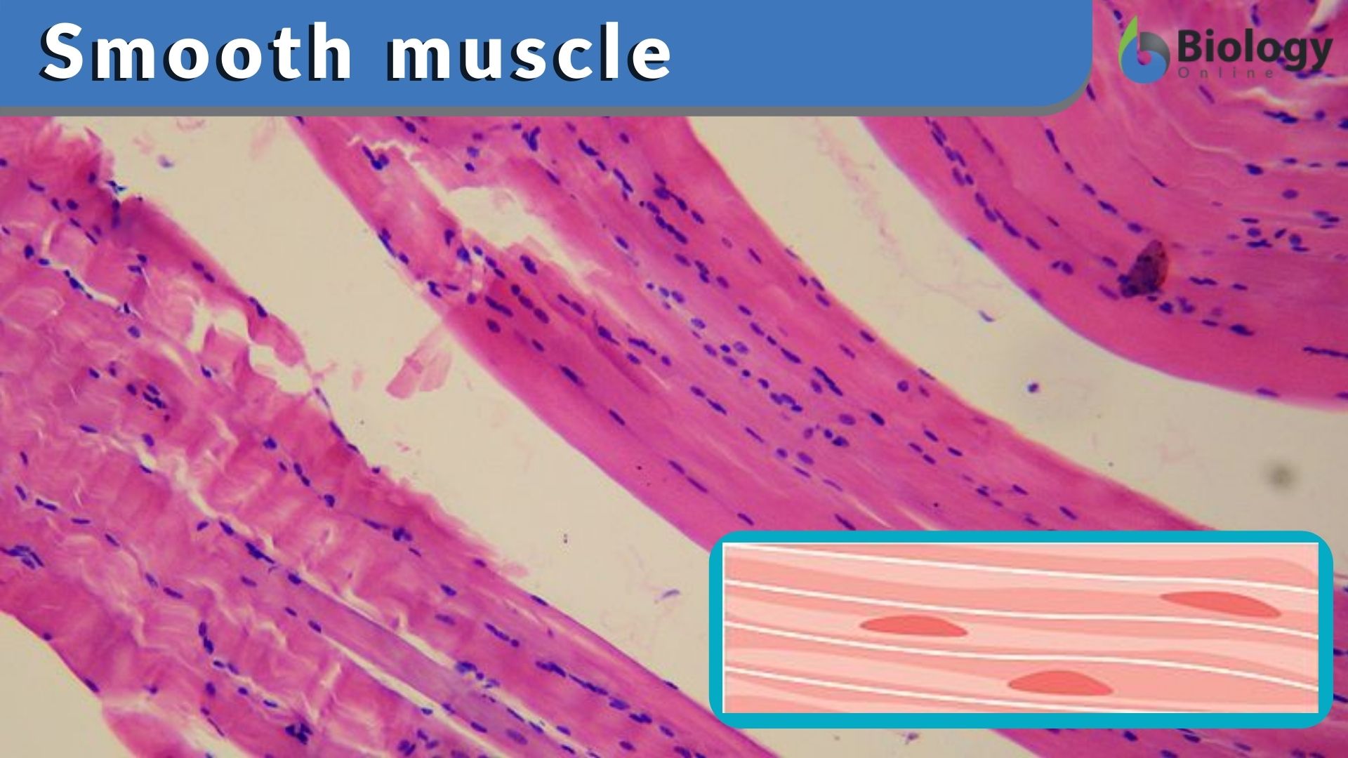
Light microscope smooth muscle endomysium.
Small amount of connective tissue surrounding each cell. Types of muscle tissue under microscope. In this scanning electron microscope image (approximately 400x) you can see the surface of a skeletal muscle fiber. Graphic design element for infographic poster, educational book or flyer. Light microscope smooth muscle identification problems. There are 3 different types of muscle: Professor susan anderson helps you recognise and understand the similarities and differences between the microscopic appearances of skeletal, cardiac and smooth muscle. Microscope slides of muscle tissue; Flat vector illustration isolated on white background. Download a free preview or high quality adobe illustrator ai, eps, pdf and high resolution jpeg. This site contains information about smooth muscle diagram labeled. Smooth muscle (also known as visceral muscle due to the locations in which they are present ) is one of the three main types of muscle tissue that exist in the human body. Fibers of smooth muscle group in branching bundles, which allows for cells to contract much stronger than those of striated musculature.
Fibers of smooth muscle group in branching bundles, which allows for cells to contract much stronger than those of striated musculature. Types of muscle tissue under microscope. In this scanning electron microscope image (approximately 400x) you can see the surface of a skeletal muscle fiber. Smooth muscle tissue, unlike striated muscle, contracts slowly and automatically. Colored pencils part 1 continue your study of muscle by examining the visceral (smooth) muscle under low part 1:
Muscle Physiology Muscle Types Contraction Lecturio from blog.lecturio.com It is divided into two subgroups; Find the perfect smooth muscle microscope stock illustrations from getty images. In this scanning electron microscope image (approximately 400x) you can see the surface of a skeletal muscle fiber. Cardiac muscle action potential diagram explained. The smith, a mighty man is he with large and sinewy hands. Colored pencils part 1 continue your study of muscle by examining the visceral (smooth) muscle under low part 1: Microscopic structure of muscle diagrams label the type of muscle (either skeletal, cardiac, or visceral) pictured. Small amount of connective tissue surrounding each cell.
Anatomy u0026 physiology chapter 9 part a lecture muscles.
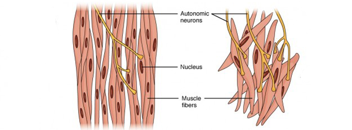
Microscopic structure of muscle diagrams label the type of muscle (either skeletal, cardiac, or visceral) pictured. Smooth muscle is under involuntary control and is innervated by the autonomic nervous system. Types of muscle tissue under microscope. Microscopic section of human kidney tissue. Professor susan anderson helps you recognise and understand the similarities and differences between the microscopic appearances of skeletal, cardiac and smooth muscle. It is divided into two subgroups; Smooth muscle has a fusiform shape, which resembles a football or spindle. Vascular smooth muscle cells(vsmc) are the major cell type in the tunica media of the aorta. The smith, a mighty man is he with large and sinewy hands. This is different from cardiac muscle tissue, which develops into an as you look at this diagram of a smooth muscle fiber, you'll notice the single nucleus in the center. Microscopic anatomy and organization of muscle tissue. Smooth muscle is found in the wall of hollow organs, passageways, tracts, eye and skin. Light microscope smooth muscle endomysium.
Smooth muscle is found in the wall of hollow organs, passageways, tracts, eye and skin. Microscopic anatomy and organization of muscle tissue. As indicated by its name, the tissue displays no striations or other distinct patterns under the microscope. Vascular smooth muscle cells(vsmc) are the major cell type in the tunica media of the aorta. Some muscles (skeletal muscles) will not contract unless stimulated by neurons;
What Are Three Types Of Muscle Cells Quora from qph.fs.quoracdn.net It is divided into two subgroups; Microscopic structure of muscle diagrams label the type of muscle (either skeletal, cardiac, or visceral) pictured. Download a free preview or high quality adobe illustrator ai, eps, pdf and high resolution jpeg. The trichome stain can be used to highlight smooth muscle cells (red) and background collagen (blue) in cases of spindled cell tumors. The smith, a mighty man is he with large and sinewy hands. Vascular smooth muscle cells (vsmcs) are the stromal cells of the vascular wall and are responsible for regulating arterial tone, blood pressure, and blood supply of the tissues. Smooth muscle (also known as visceral muscle due to the locations in which they are present ) is one of the three main types of muscle tissue that exist in the human body. Find the perfect smooth muscle microscope stock illustrations from getty images.
Smooth muscle is found in the wall of hollow organs, passageways, tracts, eye and skin.
Professor susan anderson helps you recognise and understand the similarities and differences between the microscopic appearances of skeletal, cardiac and smooth muscle. The trichome stain can be used to highlight smooth muscle cells (red) and background collagen (blue) in cases of spindled cell tumors. As indicated by its name, the tissue displays no striations or other distinct patterns under the microscope. Fibers of smooth muscle group in branching bundles, which allows for cells to contract much stronger than those of striated musculature. Compared to skeletal muscle, smooth muscle cells are small. Anatomy u0026 physiology chapter 9 part a lecture muscles. Smooth muscle is under involuntary control and is innervated by the autonomic nervous system. The muscle tissue in mammals and other higher animals is usually described as either striated, cardiac, or smooth depending on its function and appearance. Icon of smooth muscle cell under microscope. Histology of human compact bone tissue under microscope view for. Winter jasmine leaf under a microscope (leaf of winter jasmine c. Instead, they have bundles of thin and have you noticed that when you look at something under a microscope it can be very confusing, but once you look at a reference diagram or picture. Muscular tissue skeletal smooth and cardiac muscle.


0 Komentar